Essential Tests During Pregnancy
Pregnancy is one of the most important phases of a woman’s life. Physically, emotionally and cognitively different from a normal way of life, pregnancy places unique demands on the woman. Strategies are required in order to ensure a favorable outcome. This warrant tests to assure a healthy and positive outcome in pregnancy for both the mother and the fetus. Pregnancy is divided into three trimesters. Each trimester asks for different types of tests:
First-Trimester Tests
The first trimester has its own challenges. This is the phase when the fertilized egg develops into a fetus and organogenesis is active and fast paced. Any negative event may lead to birth defects in the fetus, directly impacting the outcome of pregnancy. In order to investigate how the fetus is developing, expecting mothers undergo a fetal ultrasound and a blood test. These are also called prenatal screening tests and are very effective when mixed with other tests, like Nuchal Translucency test (NT).
The first trimester has its own challenges. This is the phase when the fertilized egg develops into a fetus and organogenesis is active and fast paced. Any negative event may lead to birth defects in the fetus, directly impacting the outcome of pregnancy. In order to investigate how the fetus is developing, expecting mothers undergo a fetal ultrasound and a blood test. These are also called prenatal screening tests and are very effective when mixed with other tests, like Nuchal Translucency test (NT).
Nuchal translucency test is an ultrasound test used to determine the Nuchal thickening or increased fluid (this is the area at the back of fetal neck). Maternal blood tests are done to find the levels of human chorionic gonadotrophin (hCG) and pregnancy-associated plasma protein screening (PAPP-A). Both these substances are produced by the placenta in the first trimester. If their levels are above the normal levels, the chances of chromosomal birth defects in the fetus are high. Examples of these birth defects could be Down’s Syndrome (Trisomy 21) and Edwards’ Syndrome (Trisomy 18). In the case of detection of chromosomal defects, you must consult your obstetrician or genetic counselor for the next course of action. Additional tests like amniocentesis, chorionic villus sampling, and other ultrasounds are required for gaining a precise knowledge about the birth defect
Second-Trimester Tests
Between the 15th and 20th week of pregnancy, the blood sample from the mother is taken to investigate for multiple markers, which include the following:
Between the 15th and 20th week of pregnancy, the blood sample from the mother is taken to investigate for multiple markers, which include the following:
- Alpha-fetoprotein (AFP)
- hCG (human chorionic gonadotrophin)
- Estriol
- Inhibin.
Though not conclusive or diagnostic, these markers are indicative of birth defects in the fetus. hCG (human chorionic gonadotropin), Estriol, and Inhibin are produced by the placenta, while Alpha-fetoprotein (AFP) is produced by the fetal liver. The ideal time to look for these markers is between the 16th to 18th week of pregnancy. Alpha-fetoprotein (AFP) is synthesized by the fetal liver and is present in the amniotic fluid (this fluid surrounds the fetus). It crosses the placenta into the maternal blood circulation. This is usually detected in the serum which gives it the name MSAFP (maternal serum AFP).
In the case of abnormal AFP levels, an ultrasound is required to look for birth defects, mainly in the spinal cord. Abnormal levels of AFP are associated with neural tube defects, Down’s syndrome, abdominal wall defects, and chromosomal abnormalities. A consultation with the physician and genetic counselor is mandatory in case of the presence of any anomalies.
Third-Trimester Tests
Usually, an ultrasound is enough if there are no complications found in the first and second trimesters. This is necessary to monitor the fetal growth and other parameters. But the presence of any signs of birth defects demands higher tests. The third-trimester ultrasound usually evaluates whether the growth of the fetus corresponds to the gestational age of the fetus. Expected date of delivery can also be determined using this scan.
Usually, an ultrasound is enough if there are no complications found in the first and second trimesters. This is necessary to monitor the fetal growth and other parameters. But the presence of any signs of birth defects demands higher tests. The third-trimester ultrasound usually evaluates whether the growth of the fetus corresponds to the gestational age of the fetus. Expected date of delivery can also be determined using this scan.
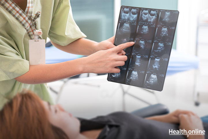
Ultrasound Scan – The Gold Standard Test in Pregnancy
Of all the tests, the ultrasound is the gold standard in pregnancy. It is mandatory to do an ultrasound scan in all the three trimesters for different purposes. But what does an ultrasound do?
Of all the tests, the ultrasound is the gold standard in pregnancy. It is mandatory to do an ultrasound scan in all the three trimesters for different purposes. But what does an ultrasound do?
An ultrasound comprises of high-frequency sound waves that are used to view the internal organs. The ultrasound can be abdominal or transvaginal. An abdominal scan visualizes the internal organs via the abdominal wall, whereas a probe is inserted into the vagina in the transvaginal ultrasound. In an abdominal ultrasound, the gel is applied to the anterior abdominal wall, whereas a condom is placed over the probe in the case of transvaginal ultrasound. The most common ultrasound generates 2D images, which can also be recorded as a video.
In the first trimester, an ultrasound confirms pregnancy, identifies the number of fetuses, locates the placenta, pinpoints the position of the fetus, investigates if there’s an ectopic pregnancy, determines if there are any fetal abnormalities, and visualizes the uterus and other support structures. The dates of the pregnancy are also determined using an ultrasound, which can be corroborated with the last menstrual period dates. The second-trimester scan is used to see if the fetus is developing appropriately for gestational age, to see the presence of neural tube defects and placental abnormalities, to observe the amniotic fluid volume, to monitor fetal growth and behavior, to oversee blood flow patterns, to measure the length of cervix, and to aid in amniocentesis. The third-trimester scan monitors fetal growth, checks the position of placenta and fetus and their relation to each other, and observes the volume of amniotic fluid.
This helps to decide and plan the mode of childbirth – if it needs to be normal or caesarean. If the placental position blocks the birth canal, then the mother may have to opt for a caesarean section.
If you are pregnant, then these tests are mandatory to monitor the fetal growth and to act early in case of any abnormal findings. Consult your obstetrician and follow her / his instructions. Regular check-ups coupled with these tests are essential for a healthy outcome in the pregnancy.