Diagnosis and Medication of Melanoma!
Many individuals go through life without ever making an appointment with a dermatologist. However, if skin cancer runs in your family, if you enjoy excessive tanning or exposure to UV rays, or if you have a lot of moles or birthmarks that have changed in color, shape, and size; you should consider conducting your own at-home skin exams, as well as talking to your doctor about getting periodic screenings from your doctor, or dermatologist. If you find a mole or marking that has changed in size, shape, color, and texture; or if the mole is bleeding, oozing, or crusting; you should consult a doctor right away.
Conducting a self-skin exam
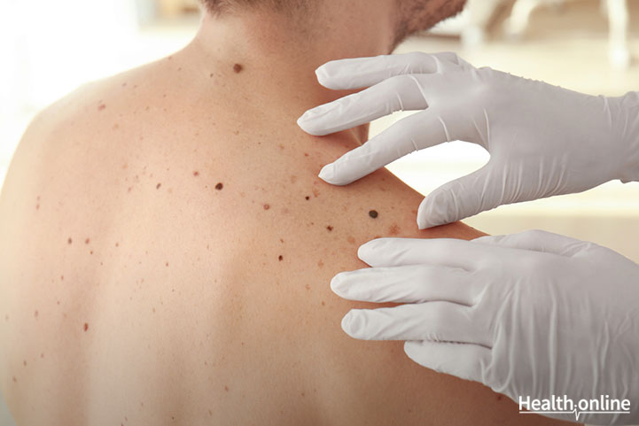
A routine skin exam is a head-to-toe inspection of your skin, paying particular attention to any skin markings, moles, freckles, or birthmarks that have changed. You can conduct your own skin exam at home. Many folks find it most convenient to conduct when they’re having a shower. If you prefer, you can also get a good look from all angles while standing in front of a full-length mirror and using a hand-held mirror for those tucked away areas. During a self-exam be sure to check your body thoroughly from head-to-toe, particularly the following:
- Scalp
- Face
- Neck
- Shoulders
- Front and back of arms
- Chest
- Torso
- Back
- Groin
- Buttocks
- Front and back of legs
- Feet
Also, don’t forget to use the hand-mirror to check in hidden away places that the sun can still reach, such as:
- Underneath fingernails and toenails
- In between fingers and toes
- Soles of the feet
You know the surface of your skin and the existing freckles and moles better than anyone else. However, if you uncover a mole or marking that looks suspicious, it’s always wise to have it looked at by a doctor, who will conduct a similar head-to-toe inspection of your skin. Your doctor may also recommend periodic skin exams or refer you to a dermatologist who specializes in skin, nails, hair health and associated conditions.
Biopsying a melanoma
According to the American Cancer Society, the only surefire way to diagnose the presence of melanoma is with a biopsy. During a biopsy, the doctor removes a sample portion of all of a suspicious skin growth or mole for laboratory analysis by a pathologist. There are various biopsies that doctors use to diagnose melanoma. Your doctor will choose the biopsy depending on the size of your mole or growth, and if only part of all of the growth needs to be removed.
Typical biopsies include:
- Shave (or tangential) biopsies are used when the risk of melanoma is considered low. The doctor will carefully shave the top layer of the skin away from the suspect area with a small surgical blade. To stop the bleeding, a topical ointment will be applied, or the doctor will use a small electrical current to cauterize the area.
- Incisional biopsies are conducted when a mole or growth is very large. The doctor will make an incision to only remove the most irregular part of a mole or growth for laboratory observation and testing.
- Punch biopsies employ a surgical tool with a circular blade that’s pressed into the skin surrounding an atypical growth or mole to remove a round sample for analysis.
- Excisional biopsies remove the whole mole or growth in addition to a portion of the surrounding border of skin for analysis.
- Optical biopsies employ a technique know as reflectance confocal microscopy (RCM), which doesn’t require extracting a skin sample.
- FNA biopsy (or fine needle aspiration) is applied to suspicious moles or large lymph nodes near a melanoma to judge if the melanoma has spread. This biopsy removes a small sample using a syringe with a thin, hollow needle and a CT scan to help guide the needle into place if the suspect area is deep in the body.
- Sentinel node biopsies are conducted to find out if the melanoma has spread to your lymph nodes by injecting a dye into the removal area and monitoring its flow into the lymph nodes. The first lymph nodes that the dye reaches are considered the sentinel lymph nodes (because they stand watch over the tumor). These are removed and tested for the presence of cancer cells. If these lymph nodes test non-cancerous, there’s good chance the melanoma has not spread.
- Surgical (or excisional) lymph node biopsies are used to remove enlarged lymph nodes if it’s suspected the melanoma has spread. This biopsy employs a small incision to get to large lymph nodes just under the skin. In this case, a local anesthetic is used; but if lymph nodes are deeper, a sedative is typically used.


