Risk Factors and Symptoms of Osteoarthritis
Risk Factors for Osteoarthritis
Osteoarthritis has several risk factors that can be broken down into three categories: modifiable local risk factors, modifiable systemic risk factors, and non-modifiable systemic risk factors.
Modifiable local risk factors are factors that occur near the site of OA that can be altered. These include muscle strength, physical activity, joint injury, joint alignment and leg length in equitability.
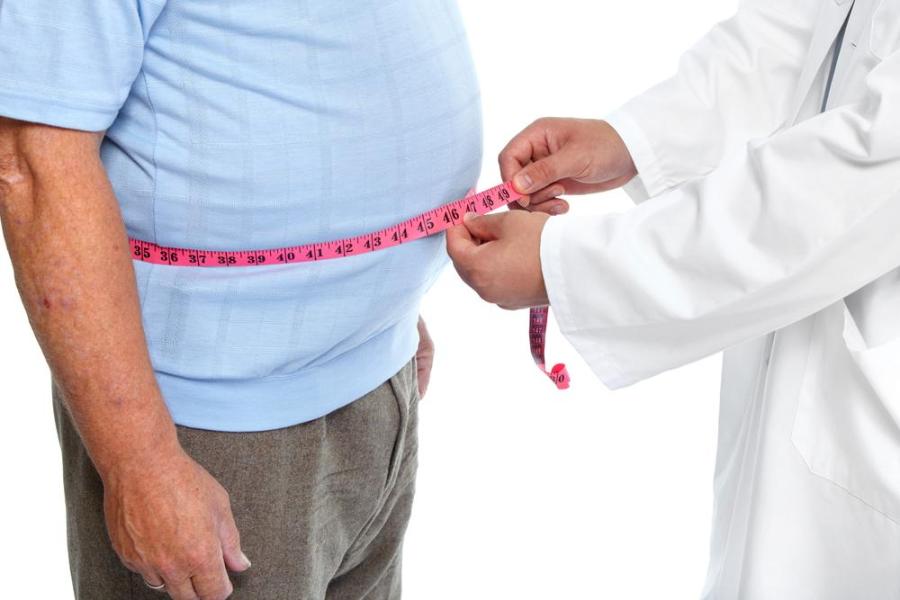
Modifiable systemic risk factors occur on a larger scale and can also be modified such as obesity and diet.
Non-modifiable system risk factors are factors that influence OA risk but cannot be changed. These include age, sex, genetics, and ethnicity.
1) Age
One of the greatest risk factors is age, which makes sense as years of wear on the bones leads to the breakdown of joint integrity. OA diagnosis increases in women especially between the ages of 60 and 64. This increase in risk with age is thought to be due to a variety of age-related changes such as a reduced capacity for joint tissue to adjust to biomechanical challenges, increased bone turnover, and accumulation of reactive oxygen that can damage articular tissue (tissue that covers the ends of the bones where they meet).
2) Gender
Women are at a greater risk for OA and typically experience more severe forms of the disease. OA in women typically affects the hand, foot, and knee most often. Severity is particularly severe after menopause, suggesting a relationship between estrogen and OA. Other reasons for these gender differences may be the difference in bone strength, alignment, ligament looseness, knee cartilage volume and neuromuscular strength as well as pregnancy.
3) Genetics
There is a large genetic component to OA. More than half of hip, hand, and knee OA have been linked to genetic factors. Several genes have been identified as contributors to OA development and maintenance by contributing to OA pathophysiological pathways.
- Chromosomes 2q has been linked to knee OA
- Chromosome 11q and chromosome 7q22 have been linked to hip OA in women and knee OA
OA in younger adults is typically caused by a previous join injury, this is known as post-traumatic OA.
4) Obesity
One of the most important risk factors for OA at the knee and hip is obesity. The correlation between weight and OA was especially strong in women compared to men.
- Jiang et al (2012) found that for every 5 unit increase in body mass index, the risk for knee OA increased 35%.
- Felson et al (1992) found that a weight loss of 5kg reduced the risk for knee OA by 50%. Obesity is also linked to hand OA, which suggests that obesity may influence OA through metabolic and inflammatory systemic effects.
5) Diet
Low vitamin levels have also been found to be associated with increased OA risk. Since vitamin D is associated with many aspects of bone and cartilage health, some studies have suggested that low vitamin D may be associated with an increased risk for knee and hip OA. However, the relationship between vitamin D and OA is still controversial.
Because reactive oxygen species accumulate over time and cause damage to articular cartilage, low antioxidant vitamin levels may also contribute to an increased risk for OA. Low vitamin C, a potent antioxidant, has been linked to an increased risk for knee OA progression while high vitamin consumption reduced the progression of radiographic knee OA by 3 fold.
Low plasma levels of vitamin K, a regulator of bone and cartilage mineralization, has been associated with increased prevalence for OA, abnormal bone protrusions (osteophytes) and joint space narrowing in the hands and osteophytes in the knees (Neogi, Booth, Zhang, Jacques, Terkeltaubl, & Aliabadi, 2006).
6) Physical Activity and Occupational Hazard
How much the joint is used may also influence the risk for osteoarthritis. Repetitive movements of the joint increase the risk for OA development. This is especially a concern for people with occupations that require continuous squatting or kneeling as they are twice as likely to develop knee OA compared to those that do not have to do these tasks. Hip OA is associated with long periods of standing or lifting while jobs that require fine hand movements have been associated with some aspects of hand OA. Proper ergonomics at work and playing sports are important to reduce risk of OA.
Expectedly, many athletes are at a higher risk of developing OA since many sports involve repetitive, high impact movements. This increased risk may also be due to increased injuries to the joint as well. Surprisingly, despite the relationship between repetitive movements and OA, an association between running and hip and knee OA has not been found (Hansen, English, & Willick, 2012).
7) Post-traumatic knee injuries
There is a strong link between joint injuries and OA. The knee is the most commonly injured joints. Those most strongly associated with OA development include an anterior cruciate ligament (ACL) tear, traumatic meniscal tears and articular cartilage damage sustained during injury. ACL ruptures involve with tremendous damage to the joint including damage to the articular cartilage, subchondral bone and collateral ligaments. Post-traumatic OA in these patients can show up as soon as 10 years after injury (Slauterbeck, Kousa, & Clifton, 2009; Lohmander, Englund, Dahl, & Roos, 2007). Thus, people that have had a joint injury are more likely to develop early onset-OA.
Joint injuries are thought to increase the risk for developing OA because the impact needed to injure the joint causes massive tissue damage that is further enhanced by the inflammation experienced by the tissue after injury. Another hypothesis is that the dysfunction of the ACL changes in the alignment of the knee which may cause regions of the bone to bear weight and interact that did not originally perform these functions (Chaudhari, Briant, Bevill, koo, & Andriacchi, 2008).
8) Muscle Weakness
A few of the many functions of muscles surrounding joints is to absorb limb loading and provide joint stability. Thus, muscle weakness may also contribute to an increased risk for OA. People with OA typically experience deficits in muscle strength and balance. There is also evidence that muscle weakness increases the chances of early onset and progression of knee OA (Bennell, Wrigley, Hunt, Lim, & Hinman, 2013). Furthermore, a study found that for every 5kg of increased extensor strength the chances of developing radiographic knee OA and symptomatic knee OA reduced by 20% and 29%, respectively (Slemenda, Brandt, Heilman, Mazzuca, Braunstein, & Katz, 1997).
9) Alignment
Incorrect joint alignment can cause an uneven distribution of load across the joint. Knee misalignment is a major predictor of knee OA progression. OA associated conditions such as bone marrow lesions and rapid cartilage loss are also associated with alignment.
Symptoms of Osteoarthritis
OA symptoms arise relatively slowly and are likely to become more severe with time. Common symptoms of OA include
- Pain during or shortly after joint movement
- Achiness
- Tenderness or sensitivity to touch
- Stiffness that occurs after an extended period of disuse such as after sleeping
- Loss of the full range of motion
- Grating sensation or cracking sound during joint movement
- Bone spurs or hard lumps of bone that form around the joint
- Swelling of joint tissue
Different symptoms may be experienced at different levels based on the OA location. Patients with OA of the hips experience pain in their groin and buttocks. Occasionally they may also experience pain on the inside of their knee or thigh. OA of the knee is typically associated with a scraping or grating sensation during movement. OA in the hands is often accompanied by bone spurs, swelling, and redness around the finger joints as well as pain at the base of the thumb. Patients with OA of the feet often experience pain at the base of their big toe or swelling around the ankles. These symptoms often result in a reduction in patient mobility. Patients with OA are also more susceptible to falls and bone fractures


